Comparison of low and high LET radiation effects in microdosimetry and dosimetry
- 17/آبان/1402
- مقالات کنفرانسی انگلیسی
- نویسنده : آزمایشگاه میکرودزیمتری نوترون
- 16 بازدید
- 0 نظر

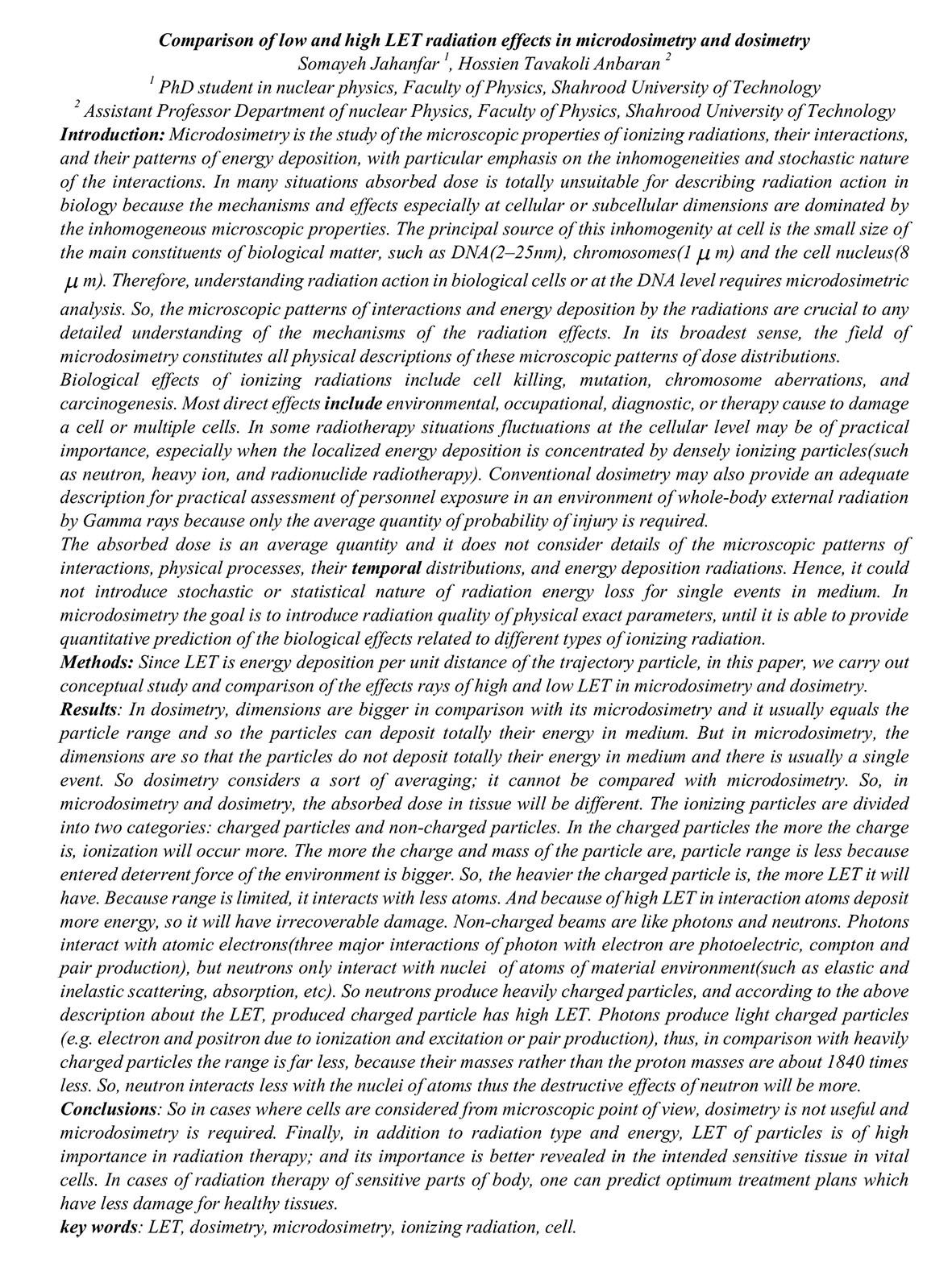
Introduction: Microdosimetry is the study of the microscopic properties of ionizing radiations, their interactions,
and their patterns of energy deposition, with particular emphasis on the inhomogeneities and stochastic nature
of the interactions. In many situations absorbed dose is totally unsuitable for describing radiation action in
biology because the mechanisms and effects especially at cellular or subcellular dimensions are dominated by
the inhomogeneous microscopic properties. The principal source of this inhomogenity at cell is the small size of
the main constituents of biological matter, such as DNA(2–25nm), chromosomes(1
m) and the cell nucleus(8 m). Therefore, understanding radiation action in biological cells or at the DNA level requires microdosimetric
analysis. So, the microscopic patterns of interactions and energy deposition by the radiations are crucial to any
detailed understanding of the mechanisms of the radiation effects. In its broadest sense, the field of
microdosimetry constitutes all physical descriptions of these microscopic patterns of dose distributions.
Biological effects of ionizing radiations include cell killing, mutation, chromosome aberrations, and
carcinogenesis. Most direct effects include environmental, occupational, diagnostic, or therapy cause to damage
a cell or multiple cells. In some radiotherapy situations fluctuations at the cellular level may be of practical
importance, especially when the localized energy deposition is concentrated by densely ionizing particles(such
as neutron, heavy ion, and radionuclide radiotherapy). Conventional dosimetry may also provide an adequate
description for practical assessment of personnel exposure in an environment of whole-body external radiation
by Gamma rays because only the average quantity of probability of injury is required.
The absorbed dose is an average quantity and it does not consider details of the microscopic patterns of
interactions, physical processes, their temporal distributions, and energy deposition radiations. Hence, it could
not introduce stochastic or statistical nature of radiation energy loss for single events in medium. In
microdosimetry the goal is to introduce radiation quality of physical exact parameters, until it is able to provide
quantitative prediction of the biological effects related to different types of ionizing radiation.
Methods: Since LET is energy deposition per unit distance of the trajectory particle, in this paper, we carry out
conceptual study and comparison of the effects rays of high and low LET in microdosimetry and dosimetry.
Results: In dosimetry, dimensions are bigger in comparison with its microdosimetry and it usually equals the
particle range and so the particles can deposit totally their energy in medium. But in microdosimetry, the
dimensions are so that the particles do not deposit totally their energy in medium and there is usually a single
event. So dosimetry considers a sort of averaging; it cannot be compared with microdosimetry. So, in
microdosimetry and dosimetry, the absorbed dose in tissue will be different. The ionizing particles are divided
into two categories: charged particles and non-charged particles. In the charged particles the more the charge
is, ionization will occur more. The more the charge and mass of the particle are, particle range is less because
entered deterrent force of the environment is bigger. So, the heavier the charged particle is, the more LET it will
have. Because range is limited, it interacts with less atoms. And because of high LET in interaction atoms deposit
more energy, so it will have irrecoverable damage. Non-charged beams are like photons and neutrons. Photons
interact with atomic electrons(three major interactions of photon with electron are photoelectric, compton and
pair production), but neutrons only interact with nuclei of atoms of material environment(such as elastic and
inelastic scattering, absorption, etc). So neutrons produce heavily charged particles, and according to the above
description about the LET, produced charged particle has high LET. Photons produce light charged particles
(e.g. electron and positron due to ionization and excitation or pair production), thus, in comparison with heavily
charged particles the range is far less, because their masses rather than the proton masses are about 1840 times
less. So, neutron interacts less with the nuclei of atoms thus the destructive effects of neutron will be more.
Conclusions: So in cases where cells are considered from microscopic point of view, dosimetry is not useful and
microdosimetry is required. Finally, in addition to radiation type and energy, LET of particles is of high
importance in radiation therapy; and its importance is better revealed in the intended sensitive tissue in vital
cells. In cases of radiation therapy of sensitive parts of body, one can predict optimum treatment plans which
have less damage for healthy tissues.


نظرات یک دیدگاه جدید ارسال کنید.
برای ثبت نظر ابتدا باید وارد سایت شوید !