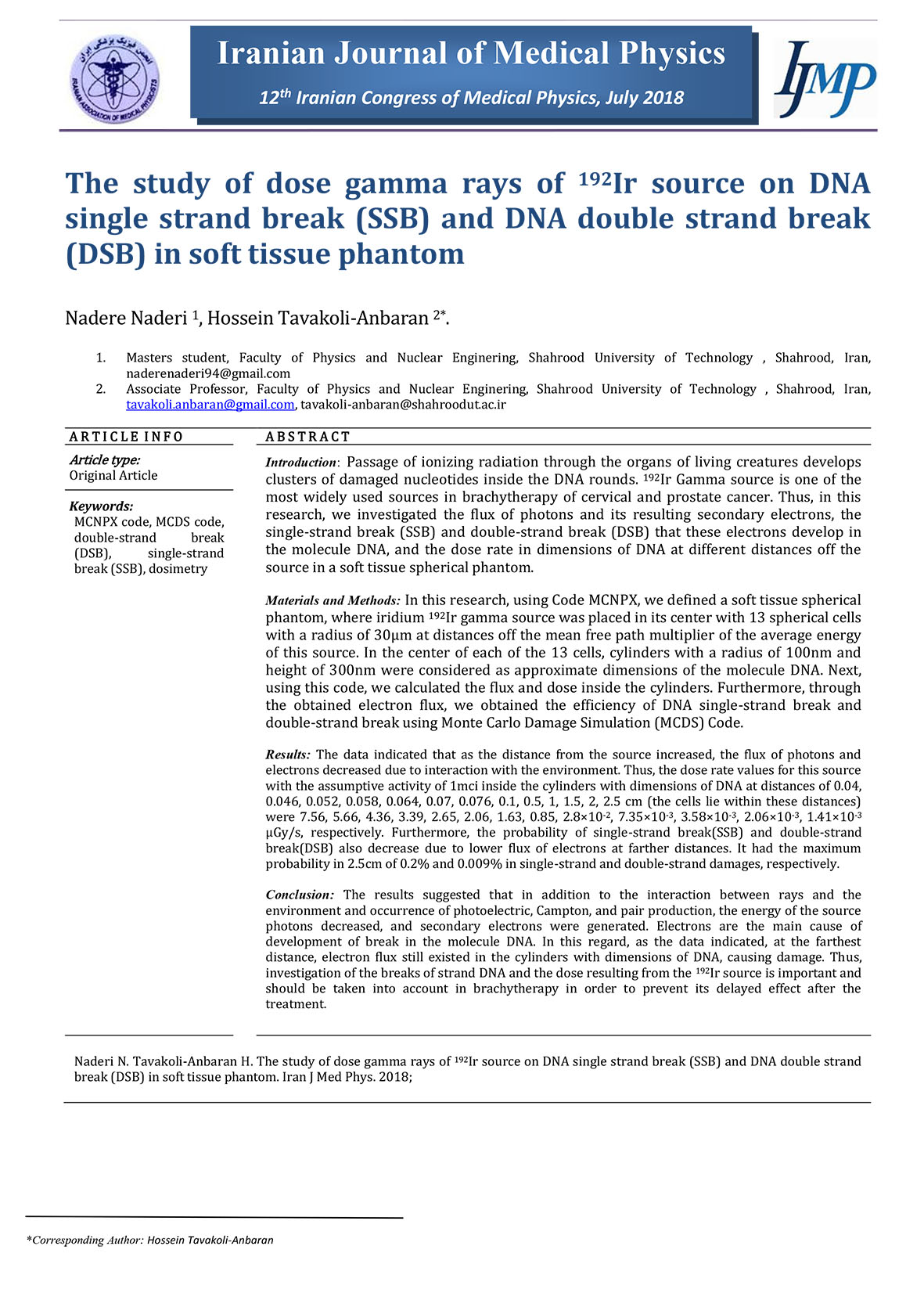
The study of dose gamma rays of 192Ir source on DNA single strand break (SSB) and DNA double strand break (DSB) in soft tissue phantom
- 17/آبان/1402
- مقالات کنفرانسی انگلیسی
- نویسنده : آزمایشگاه میکرودزیمتری نوترون
- 13 بازدید
- 0 نظر

ntroduction: Passage of ionizing radiation through the organs of living creatures develops
clusters of damaged nucleotides inside the DNA rounds. 192Ir Gamma source is one of the
most widely used sources in brachytherapy of cervical and prostate cancer. Thus, in this
research, we investigated the flux of photons and its resulting secondary electrons, the
single-strand break (SSB) and double-strand break (DSB) that these electrons develop in
the molecule DNA, and the dose rate in dimensions of DNA at different distances off the
source in a soft tissue spherical phantom.
Materials and Methods: In this research, using Code MCNPX, we defined a soft tissue spherical
phantom, where iridium 192Ir gamma source was placed in its center with 13 spherical cells
with a radius of 30μm at distances off the mean free path multiplier of the average energy
of this source. In the center of each of the 13 cells, cylinders with a radius of 100nm and
height of 300nm were considered as approximate dimensions of the molecule DNA. Next,
using this code, we calculated the flux and dose inside the cylinders. Furthermore, through
the obtained electron flux, we obtained the efficiency of DNA single-strand break and
double-strand break using Monte Carlo Damage Simulation (MCDS) Code.
Results: The data indicated that as the distance from the source increased, the flux of photons and
electrons decreased due to interaction with the environment. Thus, the dose rate values for this source
with the assumptive activity of 1mci inside the cylinders with dimensions of DNA at distances of 0.04,
0.046, 0.052, 0.058, 0.064, 0.07, 0.076, 0.1, 0.5, 1, 1.5, 2, 2.5 cm (the cells lie within these distances)
were 7.56, 5.66, 4.36, 3.39, 2.65, 2.06, 1.63, 0.85, 2.8×10⁻², 7.35×10⁻³, 3.58×10⁻³, 2.06×10⁻³, 1.41×10⁻³
μGy/s, respectively. Furthermore, the probability of single-strand break(SSB) and double-strand
break(DSB) also decrease due to lower flux of electrons at farther distances. It had the maximum
probability in 2.5cm of 0.2% and 0.009% in single-strand and double-strand damages, respectively.
Conclusion: The results suggested that in addition to the interaction between rays and the
environment and occurrence of photoelectric, Campton, and pair production, the energy of the source
photons decreased, and secondary electrons were generated. Electrons are the main cause of
development of break in the molecule DNA. In this regard, as the data indicated, at the farthest
distance, electron flux still existed in the cylinders with dimensions of DNA, causing damage. Thus,
investigation of the breaks of strand DNA and the dose resulting from the 192Ir source is important and
should be taken into account in brachytherapy in order to prevent its delayed effect after the
treatment


نظرات یک دیدگاه جدید ارسال کنید.
برای ثبت نظر ابتدا باید وارد سایت شوید !